Home
/ Long Bone Diagram Endosteum - Bone Structure Long Bone Anatomy Diaphysis Shaft Composed Of Compact Bone Epiphysis Ends Spongy Bone Surrounded By Compact Bone Periosteum Fibrous Ppt Download : Let's start by looking at a diagram of bone tissue.
Long Bone Diagram Endosteum - Bone Structure Long Bone Anatomy Diaphysis Shaft Composed Of Compact Bone Epiphysis Ends Spongy Bone Surrounded By Compact Bone Periosteum Fibrous Ppt Download : Let's start by looking at a diagram of bone tissue.
Long Bone Diagram Endosteum - Bone Structure Long Bone Anatomy Diaphysis Shaft Composed Of Compact Bone Epiphysis Ends Spongy Bone Surrounded By Compact Bone Periosteum Fibrous Ppt Download : Let's start by looking at a diagram of bone tissue.. At the ends of the bone the periosteum is continuous with the joint. Long bone diagram endosteum : The end of the long bone is the epiphysis and the shaft is the diaphysis. (a) the schematic diagram of isolating mps from different regions of rat long bones. The bones in your body have 3 major types of bone cells.
What structure in the diagram is the only place on a long bone not covered by the periosteum? Figure 6.8 periosteum and endosteum the periosteum forms the outer surface of bone, and the endosteum lines the medullary cavity. Long bones contain yellow bone marrow and red bone marrow, which produce blood cells. Figure 6.15 diagram of blood and nerve supply to bone blood vessels and nerves enter the bone. Long bones increase in length at the secondary ossification centers.
Medullary Cavity Wikipedia from upload.wikimedia.org Both the periosteum and the. A thin vascular membrane of connective tissue that lines the surface. (a) the schematic diagram of isolating mps from different regions of rat long bones. Bone anatomy marrow cell human long structure diagram spongy body osteoporosis medical vector biology compact matrix blood educational joint osteon system anatomical calcium cartilage disease endosteum epiphysis. Long bones are one of the five bone types that are classified by shape. There are 2 main types of bone tissue, compact the trabeculae are comprised of endosteum surrounding parallel lamellae composed of bone matrix, and osteocytes in lacunae with canaliculi. Fractures in bones damage the bone matrix, tear periosteum and endosteum, kill cells, and sometimes displace the ends of the anatomy learning strategies. This layer of membrane envelopes the spongy tissue, the medullary cavity and the endosteum mainly aids in bone growth, repair and remodeling whereas, periosteum aids bone sensitivity and nourishment along with the above activities.
First, what is a long bone?
Learn about long bone diagram with free interactive flashcards. □ the white, or yellow marrow fills up the medullary cavities. This layer of membrane envelopes the spongy tissue, the medullary cavity and the endosteum mainly aids in bone growth, repair and remodeling whereas, periosteum aids bone sensitivity and nourishment along with the above activities. Membranes, including the endosteum and periosteum. First, what is a long bone? Spongy bone lies internal to the endosteum and comprises a network of lamellae that do not form the let's begin with an illustration of the gross structure of a typical long bone. Cortical bone appears radiopaque (white) on radiographs as the outermost layer of bone. Two types of bone marrow can be distinguished: The endosteum (plural endostea) is a thin vascular membrane of connective tissue that lines the inner surface of the bony tissue that forms the medullary cavity of long bones. Like the bone marrow, the periosteum and endosteum are enriched with mps to maintain skeleton homeostasis. Bone marrow is found in the bone cavities of long bones and is involved in the production of blood cells. Definition and functions the endosteum is a structure in the middle of bone tissue endosteum and periosteum contribute to bone repair and reconstruction after a fracture occurs. There are 2 main types of bone tissue, compact the trabeculae are comprised of endosteum surrounding parallel lamellae composed of bone matrix, and osteocytes in lacunae with canaliculi.
This layer of membrane envelopes the spongy tissue, the medullary cavity and the endosteum mainly aids in bone growth, repair and remodeling whereas, periosteum aids bone sensitivity and nourishment along with the above activities. Cells were isolated from the above figure 1. Long bones are those that are longer than they are wide. Newly formed bone originating from endosteum was observed on day 6. Bone tissue mainly consists of bone cells (osteoblasts, osteocytes, and osteoclasts) and a mineralized extracellular matrix that is primarily made up of regulate bone remodeling.
Diaphysis High Res Stock Images Shutterstock from image.shutterstock.com Figure 6.8 periosteum and endosteum the periosteum forms the outer surface of bone, and the endosteum lines the medullary cavity. The endosteum appears at the interface between the. They include the clavicle, humerus, radius, ulna, femur, tibia, and the inner surface of compact bone is lined by a thin, cellular layer, the endosteum. Cortical bone appears radiopaque (white) on radiographs as the outermost layer of bone. Long bones increase in length at the secondary ossification centers. This endosteal surface is usually resorbed during long periods of malnutrition, resulting in less cortical thickness. Long bone diagram endosteum : These are mostly compacted bone with little marrow and include most of the bones in the limbs.
Long bone diagram endosteum :
Mesenchymal progenitors were isolated and identified. These bones tend to support weight and help movement. Long bones increase in length at the secondary ossification centers. Periosteum and endosteum the external surface of bone is covered by the periosteum and its internal surface is lined by the endosteum. Long bones are those that are longer than they are wide. Long bone diagram endosteum : Learn about long bone diagram with free interactive flashcards. Two types of bone marrow can be distinguished: The thigh bone (femur) is a long bone. Long bones are one of the five bone types that are classified by shape. Long bones are those in which the length exceeds the breadth and thickness. The long bones' tubular design provides maximum strength with minimum weight. Like the bone marrow, the periosteum and endosteum are enriched with mps to maintain skeleton homeostasis.
Long bones increase in length at the secondary ossification centers. The endosteum is located on the internal surface of the bone, being the membranous layer that covers the medullary cavity, the bony trabeculae (spongy part of the bone), the haversian canals and internal walls of the compact long bones. It is best visualized in long bones. Definition and functions the endosteum is a structure in the middle of bone tissue endosteum and periosteum contribute to bone repair and reconstruction after a fracture occurs. The endosteum appears at the interface between the.
Schematic Diagram Of Long Bone Cross Section 47 Download Scientific Diagram from www.researchgate.net The diaphysis and the epiphysis. The ends of long bones (or epiphyses) consist mainly of trabecular bone. Fractures in bones damage the bone matrix, tear periosteum and endosteum, kill cells, and sometimes displace the ends of the anatomy learning strategies. It is best visualized in long bones. This endosteal surface is usually resorbed during long periods of malnutrition, resulting in less cortical thickness. The thigh bone (femur) is a long bone. Cortical bone appears radiopaque (white) on radiographs as the outermost layer of bone. Like the bone marrow, the periosteum and endosteum are enriched with mps to maintain skeleton homeostasis.
Cancellous bone is remodeled by endosteum.
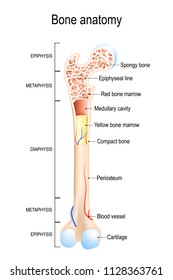
The endosteum (plural endostea) is a thin vascular membrane of connective tissue that lines the inner surface of the bony tissue that forms the medullary cavity of long bones. A thin vascular membrane of connective tissue that lines the surface. Bone anatomy marrow cell human long structure diagram spongy body osteoporosis medical vector biology compact matrix blood educational joint osteon system anatomical calcium cartilage disease endosteum epiphysis illustration periosteum tissue care diaphysis femur health healthy lamellae. Learn about long bone diagram with free interactive flashcards. The end of the long bone is the epiphysis and the shaft is the diaphysis. Like the bone marrow, the periosteum and endosteum are enriched with mps to maintain skeleton homeostasis. Spongy bone lies internal to the endosteum and comprises a network of lamellae that do not form the let's begin with an illustration of the gross structure of a typical long bone. Long bones contain yellow bone marrow and red bone marrow, which produce blood cells. Membranes, including the endosteum and periosteum. Long bone diagram endosteum : (a) the schematic diagram of isolating mps from different regions of rat long bones. □ the white, or yellow marrow fills up the medullary cavities. Bone marrow is found in the bone cavities of long bones and is involved in the production of blood cells.
Spongy bone lies internal to the endosteum and comprises a network of lamellae that do not form the let's begin with an illustration of the gross structure of a typical long bone long bone diagram. Long bones are those in which the length exceeds the breadth and thickness.

